

Overview: Inflammatory sleep disruption occurs when persistent, low-level immune activation — elevated cytokines, overactive inflammatory pathways, and immune cells in a sustained activated state — can interfere with the biological processes that produce and maintain sleep. Five mechanisms appear to contribute to it:
- Cytokine overload — chronic elevation of IL-6, TNF-α, and IL-1β is associated with more fragmented, less restorative sleep, altered REM/NREM balance, and disrupted day-night inflammatory timing
- Histamine from mast cells — brain-resident immune cells release histamine that can reinforce wake-promoting circuitry, particularly late in the sleep period when sleep pressure is lower
- Gut-to-brain inflammatory transmission — compromised intestinal barriers allow bacterial endotoxins into circulation, activating inflammatory cascades that signal to the brain through humoral routes and vagal pathways
- Neuroinflammation and impaired waste clearance — chronically activated microglia and astrocytes can disrupt the generation of deep sleep and may impair the glymphatic drainage that clears amyloid-β and tau
- Circadian clock interference — NF-κB, through its RELA/p65 subunit, can bind directly to the core circadian clock protein BMAL1, dampening clock gene expression and destabilizing sleep-wake timing
These pathways compound each other. Gut inflammation activates NF-κB, which disrupts circadian timing, which impairs deep sleep, which reduces glymphatic drainage, which allows inflammatory proteins to accumulate. Many people have more than one cause contributing to their sleep disruption.
Written by Kat Fu, M.S., M.S. · Last reviewed: December 28, 2025 · 16+ references cited
What Is Chronic Inflammation?
Acute inflammation is visible. You cut your finger, it swells, it heals. The immune response activates, does its job, and resolves. Chronic inflammation is different. There is no wound, no infection, no obvious trigger — but the immune response stays partially activated, producing a steady background of inflammatory molecules that accumulate over time.
The key players are cytokines — small proteins that immune cells use to communicate. IL-6, TNF-α, and IL-1β are among the better characterized. IL-6 coordinates immune responses across tissues and rises with sustained immune activation. TNF-α amplifies inflammatory cascades and recruits additional immune cells. IL-1β drives local inflammation and fever responses. C-reactive protein (CRP), produced by the liver in response to IL-6, is the standard blood marker for chronic inflammatory load.
At normal levels, these molecules are part of healthy immune function. At chronically elevated levels, the consequences are well documented: increased cardiovascular risk, accelerated metabolic decline, and faster cognitive aging. But chronic inflammation doesn’t only affect these areas. It also directly disrupts sleep — through five specific, identifiable mechanisms.
How Does Inflammation Disrupt Sleep?
The link between chronic inflammation and disrupted sleep is well established. A 2016 meta-analysis in Biological Psychiatry (Irwin, Olmstead, and Carroll) — pooling 72 studies with more than 50,000 participants — found that people reporting chronic sleep disturbance had elevated IL-6 (effect size 0.20) and C-reactive protein (effect size 0.12) compared to healthy sleepers. TNF-α, notably, was not associated with sleep disturbance at a statistically meaningful level in this analysis — suggesting IL-6 and CRP are the more reliable circulating markers of sleep-related inflammation. The association between disturbed sleep and inflammatory markers was specific to chronic, naturalistic sleep disruption — not acute laboratory sleep deprivation.
The evidence establishes that chronic inflammation and sleep disruption coexist.
5 mechanisms below explain how.
How Do Cytokines Fragment Sleep?
IL-1β and TNF-α are not just molecules that disrupt sleep.
At physiological levels, both participate directly in sleep regulation. IL-6 also shows sleep- and circadian-related effects, although its role in normal sleep regulation is less firmly established. IL-1β rises with prolonged wakefulness and sleep loss and promotes NREM sleep. TNF-α can enhance NREM sleep and slow-wave activity in experimental settings, although the effect depends on dose, route, timing, and species. IL-1β and TNF-α are part of the machinery that helps generate homeostatic sleep pressure.
The problem begins when they stay elevated.
Research on chronic insomnia has linked it to altered IL-6 secretion timing. In healthy sleepers, IL-6 follows a circadian rhythm that supports nighttime sleep. In chronic insomnia, this rhythm can become disrupted — with IL-6 levels elevated during the day and reduced at night (Akkaoui, Palagini, and Geoffroy, 2023). The result is the “wired but tired” experience — fatigue during waking hours alongside difficulty sleeping when IL-6 should be supporting rest.
A 2023 study in Experimental and Therapeutic Medicine (Yan et al.) used a chronic sleep fragmentation model in rats to trace where inflammation appears. Serum levels of TNF-α, IL-1β, and IL-6 were all elevated compared to controls, while the anti-inflammatory cytokine IL-10 was reduced. But the study went further: brain tissue analysis showed elevated IL-1β in the hippocampus and cortex, and elevated IL-6 in the amygdala, brainstem reticular formation, and cortex. The inflammation was not limited to the bloodstream — it reached the brain regions that regulate sleep and cognition.
A 2026 study in Behavioural Brain Research (Liu et al.), using a zebrafish model of neurodegeneration, mapped the upstream pathway: sleep deprivation activated the TLR4/MyD88/NF-κB cascade, producing the highest IL-6 and TNF-α levels among all experimental conditions. While this was an animal model, the TLR4/NF-κB pathway is conserved across species — and it points to an inflammatory cascade rather than a purely passive consequence of sleep loss.
How Does Histamine Keep the Brain Awake?
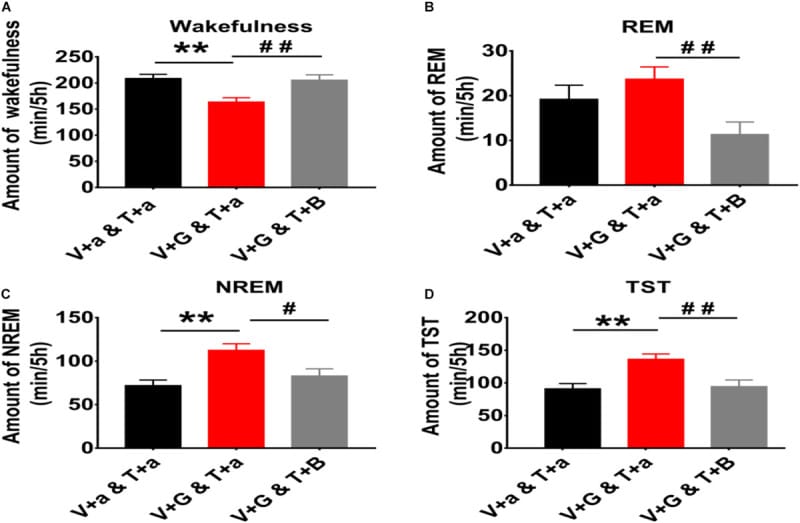
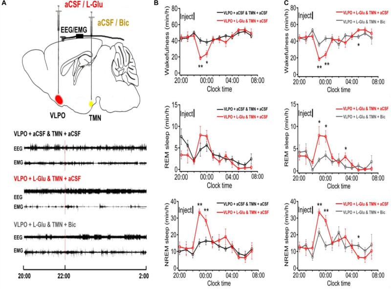
The tuberomammillary nucleus (TMN) in the posterior hypothalamus is the brain’s main wakefulness center. Its histaminergic neurons fire exclusively during waking and are suppressed during sleep. A 2021 review in the British Journal of Pharmacology (Yoshikawa, Nakamura, and Yanai) described the TMN as the control center for wakefulness — validated by the fact that pitolisant, a drug that modulates brain histamine through H3 receptors, is now approved for narcolepsy.
Sleep depends on the ventrolateral preoptic nucleus (VLPO) suppressing the TMN. The VLPO sends GABAergic inhibitory projections to TMN neurons, suppressing their histamine output during sleep. The TMN sends histamine back to suppress the VLPO during waking. This mutual inhibition creates a flip-flop mechanism: the brain is either awake or asleep, with rapid transitions between the two states. A 2020 study in Frontiers in Neuroscience (Cheng et al.) demonstrated this at the receptor pharmacology level — blocking GABA-A receptors in the TMN prevented the VLPO from inducing sleep, and blocking H1 receptors in the VLPO prevented the TMN from sustaining wakefulness.
Any factor that tonically activates the TMN tips the balance toward wakefulness.
Brain-resident mast cells are innate immune cells that release histamine and other mediators in response to immune and inflammatory signals. They are distinct from the neurons of the TMN. In mice, mast-cell-derived histamine can promote wakefulness, but in humans the dominant established wake-promoting histamine source is neuronal rather than mast-cell-derived. Histamine-related rhythms depend on the compartment being measured. TMN histaminergic neurons are wake-active and largely silent during sleep, whereas plasma histamine can rise in the early morning in some human studies and mast-cell conditions. In some people with allergic or mast-cell conditions, histamine-related reactions may worsen in the early morning and may coincide with easier late-night awakenings including 2-4am, when homeostatic sleep pressure has fallen and REM or lighter sleep are more common.
A 2025 case report in Frontiers in Sleep (Meckes and Meckes) documented this pathway in a 74-year-old male with post-viral insomnia that did not respond to zolpidem, trazodone, gabapentin, diazepam, or lemborexant (a dual orexin receptor antagonist). Oura Ring sleep scores were in the 30-40 range for ten months. Initial intervention with cyproheptadine restored sleep scores to 75+ within 24 hours, with loratadine, famotidine, and a low-histamine diet added subsequently to sustain improvement. The detail that distinguishes this case: orexin antagonists were ineffective, but antihistamine approaches succeeded, suggesting that the arousal was driven by histaminergic activation rather than orexin-mediated wakefulness.
How Does Gut Inflammation Reach the Brain?
The intestinal lining maintains tight junctions that prevent microbial products from entering the bloodstream. Gut dysbiosis, chronic stress, poor diet, and chronic sleep restriction can all compromise these junctions. When barrier integrity breaks down, lipopolysaccharide (LPS) — a component of gram-negative bacterial cell walls — crosses into circulation. This is called metabolic endotoxemia, and it activates TLR4 receptors on immune cells throughout the body, including microglia in the brain.
A 2021 study (Zhang et al.) demonstrated the causal role of the vagus nerve in this process. In mice given LPS to induce an inflammatory state, sleep deprivation amplified the inflammatory response — elevating IL-6 and TNF-α while reducing anti-inflammatory IL-10. Subdiaphragmatic vagotomy — surgically disconnecting the vagus nerve between gut and brain — abolished the amplified inflammatory cascade and the associated organ damage. Fecal microbiota transplant from sleep-deprived mice to germ-free recipients replicated the full inflammatory phenotype, establishing that the gut microbiome was driving the response through the vagal pathway.
The second route is metabolic.
Healthy gut bacteria produce short-chain fatty acids (SCFAs) — butyrate, propionate, and acetate — that suppress NF-κB activation and maintain intestinal barrier integrity. SCFAs also cross the blood-brain barrier. A 2023 review in Nutrients (Ju et al.) described how some SCFAs can reach the brain and can also modulate blood-brain barrier function and microglial activity. When SCFA-producing bacteria decline — as has been observed in people with disrupted sleep — the result is reduced anti-inflammatory signaling in the brain and altered GABAergic signaling along the gut-brain axis.
A 2025 study in Frontiers in Neurology (Yan et al.) is investigating this pathway directly — testing whether GABA-producing gut bacteria mediate insomnia through the microbiome-gut-brain axis, with fecal microbiota transplant as a causal test.
The relationship is bidirectional. Sleep disruption itself can damage gut tight junctions and reduce mucus secretion, increasing intestinal permeability and promoting further LPS translocation. Each cycle — poor sleep, increased gut permeability, more LPS, more inflammation, worse sleep.
How Does Neuroinflammation Impair Deep Sleep?
Microglia and the glymphatic drainage follow synchronized circadian rhythms — both peak during sleep. A 2024 study in IUBMB Life (Yang et al.) found that microglial morphological complexity peaks during sleep and that microglial state shapes glymphatic function in complex ways. When microglia were depleted experimentally, glymphatic function improved, but chemogenetic activation of microglia also enhanced glymphatic activity — indicating that the relationship between microglia and waste clearance is modulatory, not a binary on/off. What matters is not whether microglia are active, but what state they are in — and chronically inflamed microglia appear to modulate this relationship differently than healthy microglia do.
A 2025 review in Experimental Neurology (Yuan and Wang) traced the full loop: sleep deprivation activates microglia via NF-κB, leading to cytokine release in sleep-regulatory brain regions, disruption of glymphatic clearance, and accumulation of amyloid-β and tau. These misfolded proteins are themselves inflammatory, activating more microglia and producing more cytokines. The loop is self-reinforcing — neuroinflammation impairs the deep sleep that would resolve neuroinflammation.
Astrocytes play a parallel role.
They regulate glutamate clearance, which is essential for maintaining the excitatory-inhibitory balance during deep sleep. Under chronic inflammatory stress, astrocytic dysfunction can contribute to cortical hyperexcitability — a state incompatible with the slow oscillations that characterize restorative NREM sleep. A 2025 review in Biomedicines (Rábago-Monzón et al.) described how chronic stress specifically promotes pro-inflammatory microglial states that impair both adenosine accumulation (the molecular driver of sleep pressure) and glymphatic drainage, creating a dual deficit: the brain cannot generate adequate sleep pressure, and when sleep does occur, it cannot clear waste effectively.
The glymphatic connection has particular relevance for long-term brain health. Impaired clearance of amyloid-β and tau is linked to neurodegenerative risk — a concern that weighs heavily for adults in midlife and beyond who are already experiencing progressive inflammatory sleep disruption.
How Does Inflammation Disrupt the Body Clock?
The molecular circadian clock runs on a feedback loop driven by CLOCK and BMAL1 proteins. These proteins pair together and drive transcription of Per, Cry, and other clock genes at E-box elements in the genome. The Per and Cry proteins then accumulate and feed back to suppress CLOCK/BMAL1 activity, creating a roughly 24-hour oscillation that coordinates sleep-wake timing, hormone release, and immune function.
A 2021 study in PLoS Genetics (Shen et al.) demonstrated that the NF-κB subunit RELA binds directly to BMAL1’s transactivation domain — the same binding site used by the clock repressor CRY1 and the coactivator CBP/p300. Pharmacological and genetic activation of RELA shortened the circadian period and dampened its amplitude, while NF-κB inhibition lengthened the period. ChIP analysis showed that RELA, BMAL1, and CLOCK binding sites converge on the E-boxes of core clock genes. The implication: chronic inflammation, by sustaining NF-κB activation, progressively disrupts circadian timing from the molecular level up.
A 2022 review in ASN Neuro (Srinivasan and Walker) added a time-of-day dimension to this relationship. Glucocorticoids normally suppress NF-κB-mediated inflammation in a circadian-dependent manner. During the resting phase — when cortisol is at its lowest — NF-κB p65 expression increases, creating a window of elevated inflammatory vulnerability. This low-cortisol phase overlaps with the early morning hours when sleep is lightest and waking tends to occur. When cortisol regulation is already disrupted or when chronic inflammation has weakened circadian amplitude, this pre-dawn vulnerability widens.
A 2026 preprint (Devkar et al.) showed that sleep disruption upregulates BMAL1 in monocytes in a way that paradoxically amplifies inflammatory immune cell trafficking — monocytes from chronodisrupted mice showed biased migratory preference toward inflamed tissue, suggesting that circadian disruption doesn’t just release inflammatory brakes but actively redirects immune cell behavior.
What Triggers or Worsens Inflammatory Sleep Disruption?
How Does Chronic Stress Drive Inflammatory Sleep Disruption?
Sustained HPA activation elevates cortisol, which at high levels suppresses NF-κB activity. But the problem emerges during the troughs. When cortisol drops — particularly in the pre-dawn hours — NF-κB p65 expression rebounds, and the accumulated inflammatory priming from chronic stress expresses itself as cytokine production during the window when sleep is vulnerable to disruption.
Chronic stress also damages gut tight junctions, increasing intestinal permeability and LPS translocation into circulation. And it promotes pro-inflammatory microglial states in the brain, reducing the capacity for glymphatic clearance during whatever deep sleep remains. The result is a multi-pathway amplification: stress weakens the gut, activates brain immune cells, dysregulates cortisol timing, and fragments sleep — and fragmented sleep amplifies every one of those processes.
Does Body Composition Affect Inflammatory Sleep Disruption?
Adipose tissue is not inert storage. Visceral fat in particular produces IL-6, TNF-α at levels that can match or exceed what immune activation produces. IL-6 released from visceral adipose tissue also stimulates hepatic CRP production, which is why abdominal adiposity is so often accompanied by elevated CRP. For men in their 40s through 60s — a demographic with high rates of visceral fat accumulation — this represents a source of chronic inflammatory load that runs independent of stress, diet, or gut health.
The metabolic overlap compounds the effect. Insulin resistance — common in the same population — elevates inflammatory markers further and independently disrupts sleep architecture through cortisol and blood sugar regulation.
Does Inflammation Sensitivity Increase with Age?
The aging immune response produces what researchers call “inflammaging” — a chronic, low-grade elevation of inflammatory cytokines without acute infection. Baseline IL-6, TNF-α, and CRP rise gradually through midlife, driven by cellular senescence, mitochondrial changes, reduced immune surveillance, and visceral fat accumulation.
A 2025 randomized controlled trial in JAMA Psychiatry (Irwin, Boyle, Cho et al.) tested this relationship directly. Researchers administered an endotoxin challenge (a controlled inflammatory stimulus) to 160 adults aged 60-80, comparing those with insomnia to those without. Both groups showed similar IL-6 and TNF-α increases in response to the endotoxin. But the insomnia group showed a three-fold greater increase in depressive mood — a sustained response that reached clinically meaningful levels. The difference was not in how much inflammation the body produced. It was in how intensely the brain responded to the same inflammatory input.
This finding reframes the relationship between insomnia and inflammation in older adults. Insomnia does not only cause more inflammation — it sensitizes the brain to respond more acutely to whatever inflammation is already present. Each cycle of poor sleep increases sensitivity, and increased sensitivity makes the next cycle worse.
Circadian clock function also weakens with age, and BMAL1 expression and rhythmic amplitude decline across multiple tissues in aging models. That supports the broader point that aging can reduce circadian restraint on inflammatory signaling, but the exact magnitude is tissue-specific.
How Do You Know If Inflammation Is Disrupting Your Sleep?
Observable signs vary, but several patterns are distinctive. Waking with nasal congestion, facial puffiness, or joint stiffness can indicate overnight inflammatory activity. Histamine-related experiences — itching, flushing, or increased heart rate — that worsen in the late sleep period (2-4am) may point to mast cell activation. Sleep that feels shallow and unrestorative despite adequate duration suggests impaired slow-wave generation from neuroinflammation or cytokine disruption of sleep architecture.
The quality of early morning waking matters. People who wake from inflammatory disruption often describe feeling activated rather than groggy — alert, with a buzzing or inflamed sensation, unable to return to sleep despite being tired. This distinguishes it from the cortisol-driven waking of autonomic disruption, where the experience is often a racing mind.
Measurable markers exist. High-sensitivity C-reactive protein (hs-CRP) and IL-6 are accessible blood tests that reflect chronic inflammatory load. Fasting insulin and markers of metabolic health provide indirect evidence of inflammatory contributions. Heart rate variability patterns during sleep, now available through wearable devices, can provide physiologic context for the degree of autonomic inflammatory activation.
Many people have more than one cause contributing to their sleep disruption. Inflammation often compounds autonomic, metabolic, or hormonal disruption — and addressing one pathway can influence others. If inflammatory sleep disruption sounds like part of the picture, identifying which causes are active is a useful next step.
Find out which causes are driving your 3am wakeups →
Frequently Asked Questions
Can Inflammation Cause 3am Wakeups?
The late sleep period is where several inflammatory mechanisms can converge. Histamine-related arousal may worsen, cortisol is at its nadir, and the lighter sleep stages of the second half of the night offer less resistance to arousal. A person with elevated inflammatory histamine burden, NF-κB-driven circadian disruption, or cytokine-mediated sleep fragmentation may experience more than one simultaneously — which is why early morning waking from inflammatory causes can be resistant to standard sleep interventions.
Does Poor Sleep Make Inflammation Worse?
The Ballesio meta-analysis suggested that inflammatory changes are more likely after repeated partial sleep deprivation than after a single night. But once chronic restriction engages the inflammatory response, the bidirectional loop can help sustain itself even if the original cause of sleep disruption is addressed. Gut barrier damage from poor sleep increases LPS translocation, which activates TLR4/NF-κB, which produces cytokines that fragment sleep further. Breaking this loop typically requires addressing multiple pathways rather than a single variable.
Can You Measure Inflammatory Sleep Disruption?
No single biomarker is specific to inflammatory sleep disruption — hs-CRP and IL-6 reflect total inflammatory burden, not the amount directed at sleep-regulatory pathways. But elevated levels in someone with the characteristic sleep pattern provide supporting evidence. The combination of subjective experience and objective inflammatory markers is more informative than either alone.
Does Inflammation Affect Sleep Differently as You Age?
The aging trajectory is not a steady decline but an accelerating interaction between inflammation and sleep disruption. The Irwin 2025 trial demonstrated that the problem in older adults with insomnia is not more inflammation — it is greater sensitivity to the inflammation that is already present. This means that even modest inflammatory reductions may produce disproportionate improvements in sleep quality for older adults whose brains have become sensitized through years of chronic sleep disruption.
Related Reading
- What Is the Connection Between Chronic Inflammation and Insomnia? — The broad entry point for inflammatory sleep disruption.
- Why Does Histamine Wake You Up at 3am? — How histamine can push the brain toward wakefulness late in the night.
- Why Is Inflammation Worse at Night? — How immune timing can make sleep more vulnerable overnight.
- How Does Leaky Gut Affect Sleep? — How gut-barrier disruption can signal into inflammatory sleep pathways.
- How Does the Glymphatic System Work During Sleep? — How deep sleep and brain waste clearance connect to neuroinflammation.
- What Is Autoimmune Insomnia and Why Does It Resist Standard Sleep Approaches? — How immune conditions can create more resistant sleep disruption.
- Autonomic Sleep Disruption — How stress physiology, vagal tone, and GABA overlap with inflammation.
- Metabolic Sleep Disruption — How metabolic stress and blood sugar instability interact with inflammation.
- Circadian Sleep Disruption — How immune timing and clock timing can reinforce each other.
- Hormonal Sleep Disruption in Men — How inflammation can overlap with testosterone and cortisol pathways.
- Hormonal Sleep Disruption in Women — How menopausal hormone shifts can interact with inflammatory load.
References
Cytokines and Sleep Architecture
1. Irwin, M. R., Olmstead, R., & Carroll, J. E. (2016). Sleep disturbance, sleep duration, and inflammation: A systematic review and meta-analysis of cohort studies and experimental sleep deprivation. Biological Psychiatry, 80(1), 40-52. https://pubmed.ncbi.nlm.nih.gov/26140821/
2. Ballesio, A., Fiori, V., & Lombardo, C. (2026). Effects of experimental sleep deprivation on peripheral inflammation: An updated meta-analysis of human studies. Journal of Sleep Research, 35(1), e70099. https://pubmed.ncbi.nlm.nih.gov/40474574/
3. Akkaoui, M. A., Palagini, L., & Geoffroy, P. A. (2023). Sleep immune cross talk and insomnia. Advances in Experimental Medicine and Biology, 1411, 263-273. https://pubmed.ncbi.nlm.nih.gov/36949314/
4. Yan, D. Q., Zhang, X. P., Zhang, W. H., Deng, N., Liang, Z. T., Liu, T., Wang, G. Y., Yao, Q. W., Wang, K. K., & Tong, Z. P. (2023). Establishment of a chronic insomnia rat model of sleep fragmentation using unstable platforms surrounded by water. Experimental and Therapeutic Medicine, 25(5), 233. https://pubmed.ncbi.nlm.nih.gov/37114171/
5. Liu, T., Zhang, Y., Li, X., Liu, S., Zhu, X., Ren, Y., Han, R., Liu, K., Sun, C., Ji, X., & Li, X. (2026). Sleep deprivation-induced TLR4/MyD88/NF-kB pathway dysregulation worsens cognitive impairment in Parkinson’s disease. Behavioural Brain Research, 504, 116058. https://pubmed.ncbi.nlm.nih.gov/41577012/
6. Fiedorczuk, P., Olszewska, E., Polecka, A., Walasek, M., Mroczko, B., & Kulczynska-Przybik, A. (2023). Investigating the role of serum and plasma IL-6, IL-8, IL-10, TNF-alpha, CRP, and S100B concentrations in obstructive sleep apnea diagnosis. International Journal of Molecular Sciences, 24(18), 13875. https://pubmed.ncbi.nlm.nih.gov/37762178/
Histamine and Mast Cell Wakefulness
7. Yoshikawa, T., Nakamura, T., & Yanai, K. (2021). Histaminergic neurons in the tuberomammillary nucleus as a control centre for wakefulness. British Journal of Pharmacology, 178(4), 750-769. https://pubmed.ncbi.nlm.nih.gov/32744724/
8. Meckes, A. J., & Meckes, J. W. (2025). Case report: A case of post-viral inflammatory insomnia: Observed sleep restoration associated with histamine-targeted interventions and implications for mast cell pathways. Frontiers in Sleep, 4, 1736866. https://pubmed.ncbi.nlm.nih.gov/41640426/
9. Cheng, J., Wu, F., Zhang, M., Ding, D., Fan, S., Chen, G., Zhang, J., & Wang, L. (2020). The interaction between the ventrolateral preoptic nucleus and the tuberomammillary nucleus in regulating the sleep-wakefulness cycle. Frontiers in Neuroscience, 14, 615854. https://pubmed.ncbi.nlm.nih.gov/33381012/
10. Naganuma, F., Nakamura, T., Kuroyanagi, H., Tanaka, M., Yoshikawa, T., Yanai, K., & Okamura, N. (2021). Chemogenetic modulation of histaminergic neurons in the tuberomamillary nucleus alters territorial aggression and wakefulness. Scientific Reports, 11(1), 17935. https://pubmed.ncbi.nlm.nih.gov/34504120/
11. Maurer, J., et al. (2020). The role of preoptic area GABAergic axonal projections to tuberomammillary nucleus in sleep homeostasis [Conference abstract]. Sleep, 43(Suppl_1). [Conference abstract — not indexed in PubMed]
12. Noain, D., Buchele, F., Schreglmann, S. R., Valko, P. O., Gavrilov, Y. V., Morawska, M. M., Imbach, L. L., & Baumann, C. R. (2018). Increased sleep need and reduction of tuberomammillary histamine neurons after rodent traumatic brain injury. Journal of Neurotrauma, 35(1), 85-93. https://pubmed.ncbi.nlm.nih.gov/28762870/
13. Moline, M., et al. (2022). Effect of lemborexant on early morning awakening in subjects with severe problems with waking too early [Conference abstract]. Sleep, 45(Suppl_1). [Conference abstract — not indexed in PubMed]
Gut-Brain Inflammatory Axis
14. Zhang, Y., Xie, B., Chen, X., Zhang, J., & Yuan, S. (2021). A key role of gut microbiota-vagus nerve/spleen axis in sleep deprivation-mediated aggravation of systemic inflammation after LPS administration. Life Sciences, 265, 118736. https://pubmed.ncbi.nlm.nih.gov/33176177/
15. Yan, L., Wang, X., Liu, X., Cai, J., Zhang, Z., & Yang, B. (2025). An analysis of electroacupuncture as adjunctive treatment for insomnia: A new perspective targeting GABA-mediated microbiome-gut-brain axis. Frontiers in Neurology, 16, 1504316. https://pubmed.ncbi.nlm.nih.gov/40371088/
16. Ju, S., Shin, Y., Han, S., Kwon, J., Choi, T. G., Kang, I., & Kim, S. S. (2023). The gut-brain axis in schizophrenia: The implications of the gut microbiome and SCFA production. Nutrients, 15(20), 4391. https://pubmed.ncbi.nlm.nih.gov/37892465/
17. Miranda, C. A. M., Vasconcelos, L. M., Oliveira, C. W. S., Lopes, M. C. M., & Diogo, L. R. F. (2025). Gut-brain axis: How the gut microbiome modulates sleep quality in adults. International Health Sciences Review, 1(4), 68-72. [Not indexed in PubMed]
18. Gao, Y., & Yu, X. (2026). Acupuncture treatment for insomnia based on the microbiome-gut-brain axis theory: A review. Medicine, 105(1), e46967. https://pubmed.ncbi.nlm.nih.gov/41496086/
19. Madison, A., Duff, A., & Bailey, M. (2025). Gut barrier permeability, microbiota, and brain health. In The Oxford Handbook of the Microbiome-Gut-Brain Axis. Oxford University Press. [Book chapter — not indexed in PubMed]
20. Wang, C., Xu, Z., Wang, P., Zhang, Q., & Xiang, H. (2026). Gut-heart axis disruption and LPS translocation: Driving atrial fibrillation through inflammatory storm and fibrotic mechanisms. Frontiers in Endocrinology, 17, 1787393. https://pubmed.ncbi.nlm.nih.gov/41821743/
Neuroinflammation and Glymphatic Clearance
21. Yang, T., Tang, Y., Liu, X., Gong, S., & Yao, E. (2024). Microglia synchronizes with the circadian rhythm of the glymphatic system and modulates glymphatic system function. IUBMB Life, 76(12), 1209-1222. https://pubmed.ncbi.nlm.nih.gov/39223969/
22. Yuan, X. L., & Wang, C. Y. (2025). Sleep deprivation-induced cognitive impairment: Unraveling the role of neuroinflammation. Experimental Neurology, 394, 115419. https://pubmed.ncbi.nlm.nih.gov/40818666/
23. Rabago-Monzon, A. R., Osuna-Ramos, J. F., Armienta-Rojas, D. A., Camberos-Barraza, J., Camacho-Zamora, A., Magana-Gomez, J. A., & De la Herran-Arita, A. K. (2025). Stress-induced sleep dysregulation: The roles of astrocytes and microglia in neurodegenerative and psychiatric disorders. Biomedicines, 13(5), 1121. https://pubmed.ncbi.nlm.nih.gov/40426947/
24. Gupta, S., et al. (2025). Hypoxia affects glymphatic efficiency: Assessing glymphatic disruption in obstructive sleep apnea using near-infrared spectroscopy [Conference abstract]. Sleep, 48(Suppl_1). [Conference abstract — not indexed in PubMed]
25. Lozano, K., et al. (2025). Effects of early-adulthood sleep disruption on longitudinal spatial learning, neuroinflammation, and late-life tau pathology in the PS19 mouse model [Conference abstract]. Sleep, 48(Suppl_1). [Conference abstract — not indexed in PubMed]
26. Tanaka, M. (2026). Neurogenesis and neuroinflammation in dialogue: Mapping gaps, modulating microglia, rewiring aging. Cells, 15(1), 78. https://pubmed.ncbi.nlm.nih.gov/41511361/
NF-kB and Circadian Clock
27. Shen, Y., Endale, M., Wang, W., Morris, A. R., Francey, L. J., Harold, R. L., Hammers, D. W., Huo, Z., Partch, C. L., Hogenesch, J. B., Wu, Z. H., & Liu, A. C. (2021). NF-kB modifies the mammalian circadian clock through interaction with the core clock protein BMAL1. PLoS Genetics, 17(11), e1009933. https://pubmed.ncbi.nlm.nih.gov/34807912/
28. Srinivasan, M., & Walker, C. (2022). Circadian clock, glucocorticoids and NF-kB signaling in neuroinflammation — implicating glucocorticoid induced leucine zipper as a molecular link. ASN Neuro, 14, 17590914221120190. https://pubmed.ncbi.nlm.nih.gov/36317290/
29. Devkar, R., et al. (2026). Circadian clock protein Bmal1 drives inflammatory homing in monocytes via augmented Cxcr4 and Ccr2 axis [Preprint]. Research Square. [Preprint — not indexed in PubMed]
30. Turek, F. W., & Vitaterna, M. H. (2025). Fundamentals of the circadian clock system. In Fundamentals of Sleep and Circadian Science. Oxford University Press. [Book chapter — not indexed in PubMed]
31. Zhang, X., Viggars, M., & Esser, K. (2023). Analysis of circadian transcription activator BMAL1 and CLOCK binding targets in young and old mouse skeletal muscle [Conference abstract]. Innovation in Aging, 7(Suppl_1). [Conference abstract — not indexed in PubMed]
32. Zeng, H., Yang, X., Liao, K., Zuo, X., Liang, L., He, D., Ju, R., Wang, B., & Yuan, J. (2024). Circadian disruption reduces MUC4 expression via the clock molecule BMAL1 during dry eye development. Experimental & Molecular Medicine, 56(7), 1655-1666. https://pubmed.ncbi.nlm.nih.gov/38956298/
Inflammaging and Sensitivity
33. Irwin, M. R., Boyle, C. C., Cho, J. H., Sadeghi, N., Castillo, D., Smith, M. T., Eisenberger, N. I., & Olmstead, R. (2025). Inflammatory exposure and depression in older adults with insomnia: A randomized clinical trial. JAMA Psychiatry, 82(9), 859-867. https://pubmed.ncbi.nlm.nih.gov/40668551/
34. Miao, Y., Wang, J., Li, X., Guo, J., Ekblom, M. M., Sindi, S., Zhang, Q., & Dove, A. (2025). Poor sleep health is associated with older brain age: The role of systemic inflammation. eBioMedicine, 120, 105941. https://pubmed.ncbi.nlm.nih.gov/41033940/